Different microscopy contrasts
Our hybrid method is applicable to different types of microscopy data that provide information on fiber orientation. We have successfully demonstrated its effectiveness with Polarised Light Imaging (PLI), as well as myelin-stained histology (Gallyas Silver), and nissl-stained histology (Cresyl Violet) that were first processed using structure tensor analysis [citation]. For datasets that include both diffusion MRI and microscopy data on fiber orientations within the same tissue blocks, our hybrid method has the potential to generate hybrid orientations and enable tractography.
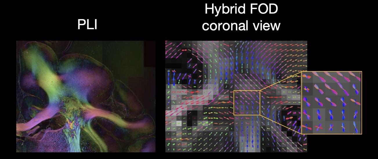
Polarised light imaging (PLI)
Polarized Light Imaging (PLI) estimates the primary fiber orientation based on the birefringence of myelinated axons, acquired with a resolution of 4 microns/pixel. Maps of fiber orientation within the microscopy plane, known as in-plane angles, were used as inputs for the hybrid dMRI-PLI tractography.
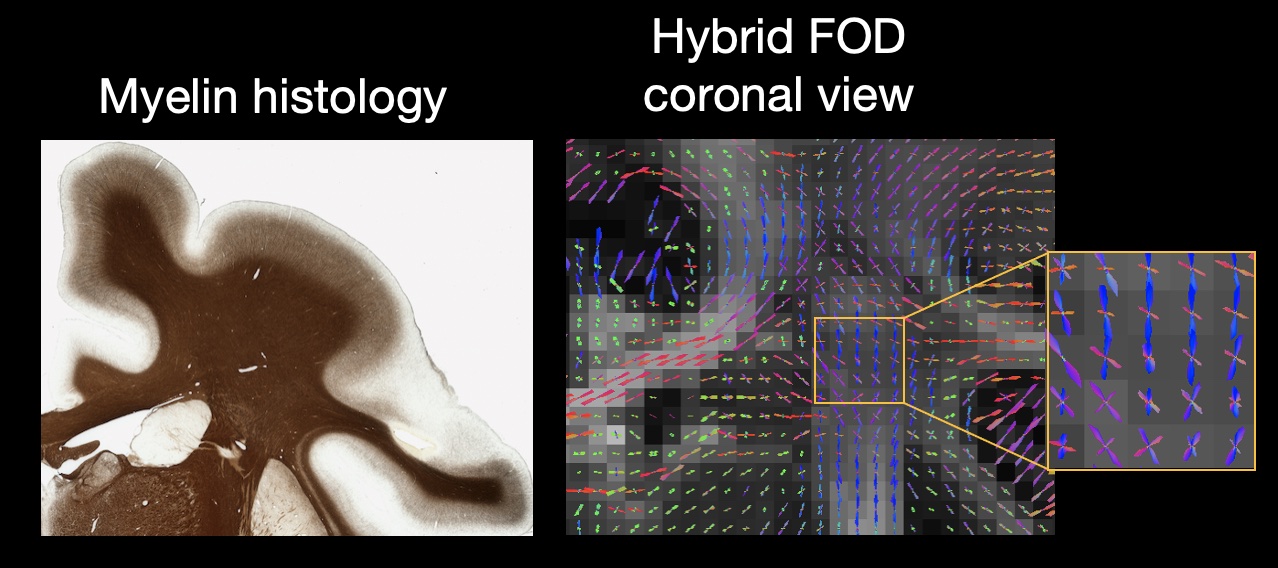
Gallyas Silver staining
The Gallyas Silver stain is a common staining method where myelin is stained brown. These slides were digitised with a resolution of 0.28 microns/pixel. Fibre orientations for each microscopy pixel were then estimated through structure tensor analysis, and the output of which was input into the hybrid method to reconstruct the hybrid Gallyas-dMRI fibre orientations.
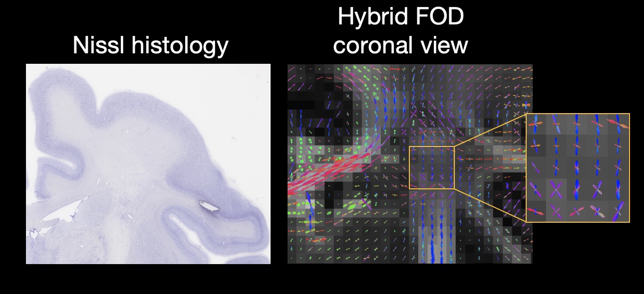
Cresyl Vilot staining
Cresyl voilet is a common Nissl-body stain where the cell bodies of both neurons and glia are stained purple. These data can again be analysed using structure tensor analysis and produce orientations generally similar to myelin-sensitive microscopy [cite]. This is due the cell bodies tending to be arranged in pattens that generally follow the underlying white matter. As above, we used the structure tensor output as input to hybrid orientation reconstruction.
Key references:
Axer, H., Axerl, M., Krings, T., Keyserlingk, D.G.v.. Quantitative estimation of 3-D fiber course in gross histological sections of the human brain using polarized light. Journal of Neuroscience Methods 2001;105(2):121{31. doi:10.1016/S0165-0270(00)00349-6.
Larsen, L., Grässel, L.D., Grässel, D., Witte, O.W., Axer, H.. Polarized light imaging of white matter architecture. Microscopy Research and Technique 2007;70(10):851-863. doi:10.1002/jemt.20488.
Axer M, Amunts K, Grässel D, Palm C, Dammers J, Axer H, Pietrzyk U, Zilles K. A novel approach to the human connectome: ultra-high resolution mapping of fiber tracts in the brain. Neuroimage 2011; 15;54(2):1091-101. doi: 10.1016/j.neuroimage.2010.08.075.
Axer, M., Grässel, D., Kleiner, M., Dammers, J., Dickscheid, T., Reckfort, J., et al. High-Resolution Fiber Tract Reconstruction in the Human Brain by Means of Three-Dimensional Polarized Light Imaging. Frontiers in Neuroinformatics 2011;5:34. doi:10.3389/fninf.2011.00034.
Gallyas, F.. Silver staining of Alzheimer’s neuro brillary changes by means of physical development. Acta Morphologica Academiae Scientiarum Hungaricae 1971;19(1):1-8.
Bigun, J., Bigun, T., Nilsson, K.. Recognition by symmetry derivatives and the generalized structure tensor. IEEE Transactions on Pattern Analysis and Machine Intelligence 2004;26(12):1590{1605. doi:10.1109/tpami.2004.126.
Budde, M.D., Frank, J.A.. Examining brain microstructure using structure tensor analysis of histological sections. NeuroImage 2012;63(1):1{10. doi:10.1016/j.neuroimage.2012.06.042.