Welcome to the dMRI-microscopy hybrid tractography tutorial
Here, we have developed a novel approach that combines diffusion MRI and microscopy for 3D white matter tract reconstruction.
Multimodal datasets combining diffusion MRI (dMRI) and microscopy afford unique opportunities for brain connectome imaging. Orientation-sensitive microscopy such as myelin-stained histology or polarised light imaging can tell us about fibre orientations in great detail due to high spatial resolution. These methods can be used to image thin tissue sections over large fields of view (e.g. whole coronal sections). However, this microscopy often provides reliable fibre orientation information only within the microscopy plane (i.e. 2D fibre orientations). This inhibits white matter tract reconstruction via tractography which requires estimation of 3D fibre orientations across the brain. Though it is possible to estimate 3D fibre orientatiions from microscopy (.e.g via 3D PLI, micro-CT or z-stack imaging), 3D microscopy is usually acquired in small tissue blocks, again limiting our ability to track fibre bundles across the brain.
In comparison, diffusion MRI is a powerful tool for in vivo white matter imaging. It provides 3D fibre orientation covering the whole brain, but has limited spatial resolution on the milimetre scale. Even at the highest achieveale resolutions, each MRI voxel contains tens of thousands of white matter fibres whose orientations are only indirectly related to the MR signal via water diffusion. Consequently, the estimation of fibre orientations from diffusion MRI can be inaccurate, resulting in downstream tractography errors such as false positive connections and/or tract reconstructions that do not follow our neuroanatomical expectations.
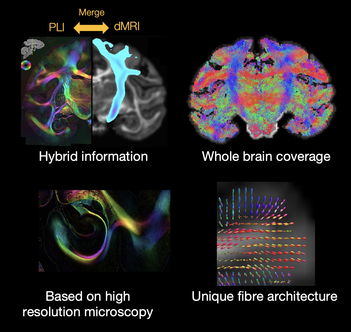
To overcome several of these limitations, we developed a hybrid method to combine the strengths of microscopy and dMRI and create “hybrid diffusion MRI-microscopy fibre orientations”. Specifically, we use 2D microscopy to provide the estimates of fibre orientation within the microscopy plane and dMRI to provide the out-of-plane orientation. We then combine the hybrid orientations over a local neighbourhood (e.g. 0.4 or 0.6 mm isotropic voxels) to create hybrid fibre orientation distributions (FODs) . These hybrid fibre orientation distributions can be input into traditional tractography pipelines to reconstruct white matter tracts across the brain or estimate the microscopy connectome.
Hybrid benefits
Our hybrid fibre orientation distributions (FODs) have the following benefits:
Our hybrid method converts 2D microscopy data into 3D fibre orientation distributions that can be input into existing tractography pipelines to perform microscopy-informed tractography.
Whole-brain coverage allows for white matter tract reconstruction across the brain or whole-brain estimates of structural connectivity.
3. Our hybrid orientations are created at the high resolution of the microscopy data and thus retain detailed information about complex fibre configurations. 5. The hybrid orientations can be combined into fibre orientation distributions (FODs) at any arbitrary resoluton, up to the native resolution of the microscopy data. This includes resolutions higher than what is typically achievable in vivo.
Tips for getting started
Want to learn more about the method? Please see the Method explanation tab.
Want to construct hybrid FODs with your own diffusion MRI and microscopy dataset? Please see the Code explanation tab.
Want to run hybrid tractography and explore its benefits? Please see the Hybrid FODs and Tractography Outputs tab.
Want to find out more about how we can create hybrid orientations from different microscopy contrasts (PLI, myelin and nissl stains)? Please see the Different microscopy contrasts tab.
Contact us
Any questions, comments, requests or ideas for collaboration, please reach out to Silei Zhu: silei.zhu@ndcn.ox.ac.uk